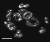


FITC Image - Telomeres;
telomere cluster is evident
RHOD Image - knobs;
direct labeled rhodamine oligo probes for knobs, NUBI-R, stains
knob sequence clusters, including small telomeric clusters.
3 Color Overlay; shows DAPI
(red), FITC-telomeres (green), and Rhodamine (white-knobs).
Scale bars are 5 micron.
Link goes to
Abstract page with links to movies, cover photo, etc.
Telomere FISH and the Bouquet Stage Images
(Murphy, Bass), maize line A344+ at the bouquet stage, as seen by
telomere FISH, from 3-D, three-wavelength image.
(Bass, Sedat, Cande), as seen by
telomere FISH, from 3-D, two-wavelength image.
(Bass, Sedat, Cande)
Same nucleus, different sections and pseudo-coloring
6sa2.JPG, Blue=DAPI,
Green=Telomere-FITC, and Red=5srDNA-Rhodamine.
6sa3.GIF, Red=DAPI,
Green=Telomere-FITC, and Blue=5srDNA-Rhodamine
(Bordoli, Bass, FSU)
Most maize lines do not reveal the bouquet structure
in DAPI-stained chromosome images. A344 + is an exception.
Here are various views (projections) of
a single nucleus at late zygotene.
DAPI Image - DNA,
the bouquet is at 11 o'clock and knobs are evident
SPIN ME
A344-bq-DAPI.mov 360 movie
SPIN ME
A344-bq-FITC.mov 360 movie
SPIN ME
A344-bq-RHOD.mov 360 movie
SPIN ME
A344-bq-color.mov 360 movieOat-Maize9 chromosome addition line MOVIES
Chromosome paint (green) and telomere FISH (purple) at meiotic prophase.
GEMINIVIRUS DNA Localized by FISH to Intranuclear Compartments

Supplemental Movies of 3D data (preview - under construction)
These data are from a collaboration with Niki Robertson and Linda
Hanley-Bowdoin at NC State University.
OTHER and for FUN
,
the GreenPower Version.
 What's wrong with this nucleus?

HOME