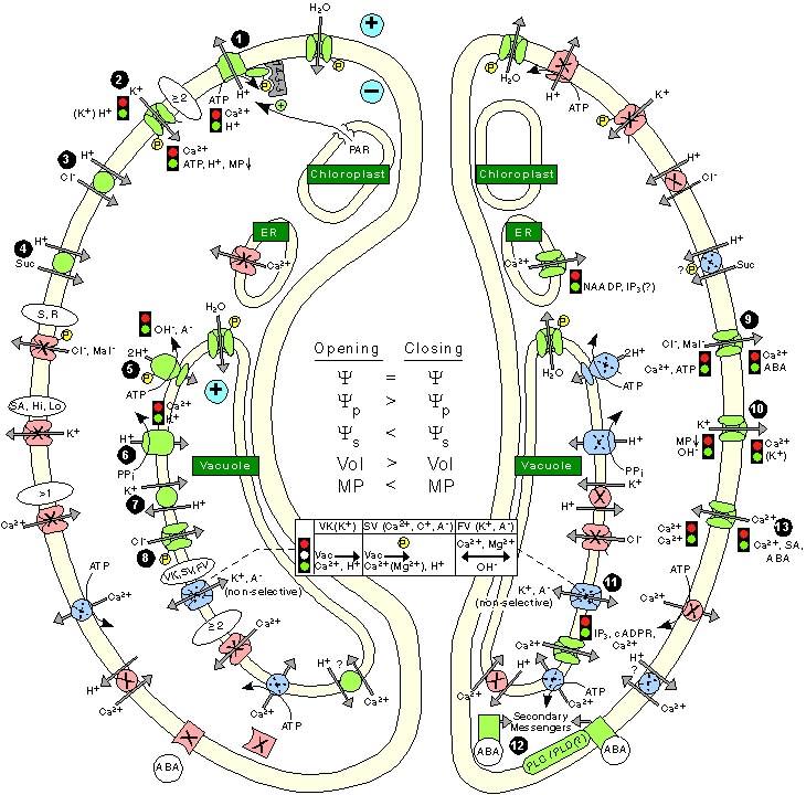
GC membrane-transport processes during stomatal opening (left) and stomatal closing (right). Transporters in a conducting state are green; in a nonconducting state, red; conducting state not specified, blue. Note, as indicated, that one icon may represent several transporters having different properties. The traffic-control icon by a transporter indicates inhibition (red, upper) or activation (green, lower); effectors act directly and indirectly. The roles of some transporters are not assigned with certainty. In essence, stomatal opening is initiated by H+ extrusion (1), which hyperpolarizes the PM and acidifies the apoplast. These effects increase the driving force for K+ uptake and activate the voltage-regulated K+-in channel (2). Cl- (3) and suc (4) may also be taken up. H+ pumping into the vacuole (5, 6) provides for K+ antiport (7) and Cl- uptake (8). GC solute increase leads to H2O uptake and therefore an increase in GC volume and aperture size. Stomatal closing is initiated by activation of the A- channel (9), which depolarizes the PM. This effect increases the driving force for K+ efflux and activates the voltage-regulated K+-out channel (10). Several channels (11) provide for solute release from the vacuole. Internal and external ABA receptors (12) cause the release of Ca2+ through cellular messengers (e.g., IP3); ABA also causes Ca2+ influx (13). ABA also induces stomatal closure by Ca2+-independent means. Compartments are not to scale.