So what was David actually doing in Villefranche?
posted 25 May 2005
We've had that question several times—what does David actually do in Villefranche—and as usual when asked the same thing at home, he finds it hard to answer. I think he has trouble telling at what scale the question is intended, and then he has trouble figuring out at what level to answer and difficulty avoiding jargon, so he hems and haws and sounds as though he doesn't want to answer. So let me take a shot, at a mixture of levels, hoping to make it clear to scientists and laymen alike.
Mornings are easy to describe. After getting up and having breakfast, we would head to the lab and get there between 8:30 and 9:00 a.m. I would install myself in a corner of Janine Cuzin's office in the Vieille Forge, spend an hour or two working on this journal and/or a free-lance project, then go on the time clock and spend the rest of the day doing pretty much what I do in Tallahassee.
David would go to his office in the Aquarium Hall, fire up his computer, and work on scientific papers. Over the last few years, he's accumulated several sets of data that, for one reason or another (most often lack of time) have never been written up for publication—side projects that were overshadowed at the time by larger experiments, data from old master's or doctoral projects that the students never developed any further, experiments whose results did not seem at the time to make sense but have since become clearer, etc. The sabbatical has given him the leisure to think about those results, consider how they might best be analyzed, do the background reading to see more clearly how they might fit into the body of existing knowledge, and actually write the papers. He's sent several off to coauthors for revision and comment and has another almost ready to go. The wall above his desk is papered in artwork by children of our acquaintance, graphs of the latest results of his project with Laurence, and graphs of the back-log dataset he's currently wrestling to interpret.
About 12:30 p.m., we would break for lunch. When the weather was warm enough, we would sit on the ponton, watching the sailboats and luxury yachts come and go while eating our sandwiches. If the weather was cold, we would eat in David's office or join those gathering in the communal kitch. After lunch, we would go for a stroll along the quais of the Port de la Darse before going back to work at about 1:30 p.m.
David would then spend some time reading the current literature (a scientist's reading is never done) before he and Laurence met in their microscopy lab to work on their joint project.
As part of a larger project that continued for several years, personnel of the Villefranche Oceanography Lab deployed sedimentation traps in very deep water in the Mediterranean—one of the advantages of the Villefranche lab is its ready access to nearby deep-sea conditions. These traps were intended to quantify, by season and year, the rain of particles that sprinkles out of the surface waters and arrives at the deep-sea bottom—dead plankton, the fine red dust that periodically blows into the area from north Africa, soil washed from the continents by runoff, etc. They were weighted and buoyed so as to be suspended about 16 feet above the bottom and were replaced on a regular schedule. To ensure that everything that fell into the traps stayed there to be tallied later, the investigators devised a clever system involving density-gradient layering of various substances that both ensured that all arriving particles dropped into a lethal and preservative formalin bath and prevented any formalin from escaping into the water. That way, organic material, like fecal pellets and dead plankton, wouldn't be, e.g., consumed by bacteria or eaten by passing fish before the trap could be retrieved and taken back to the lab.
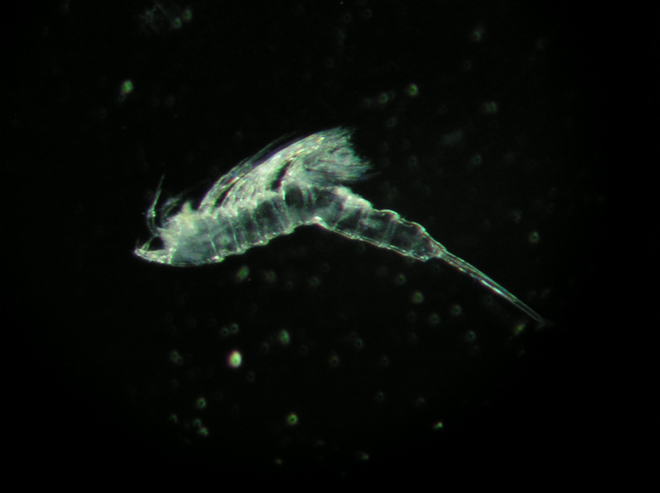
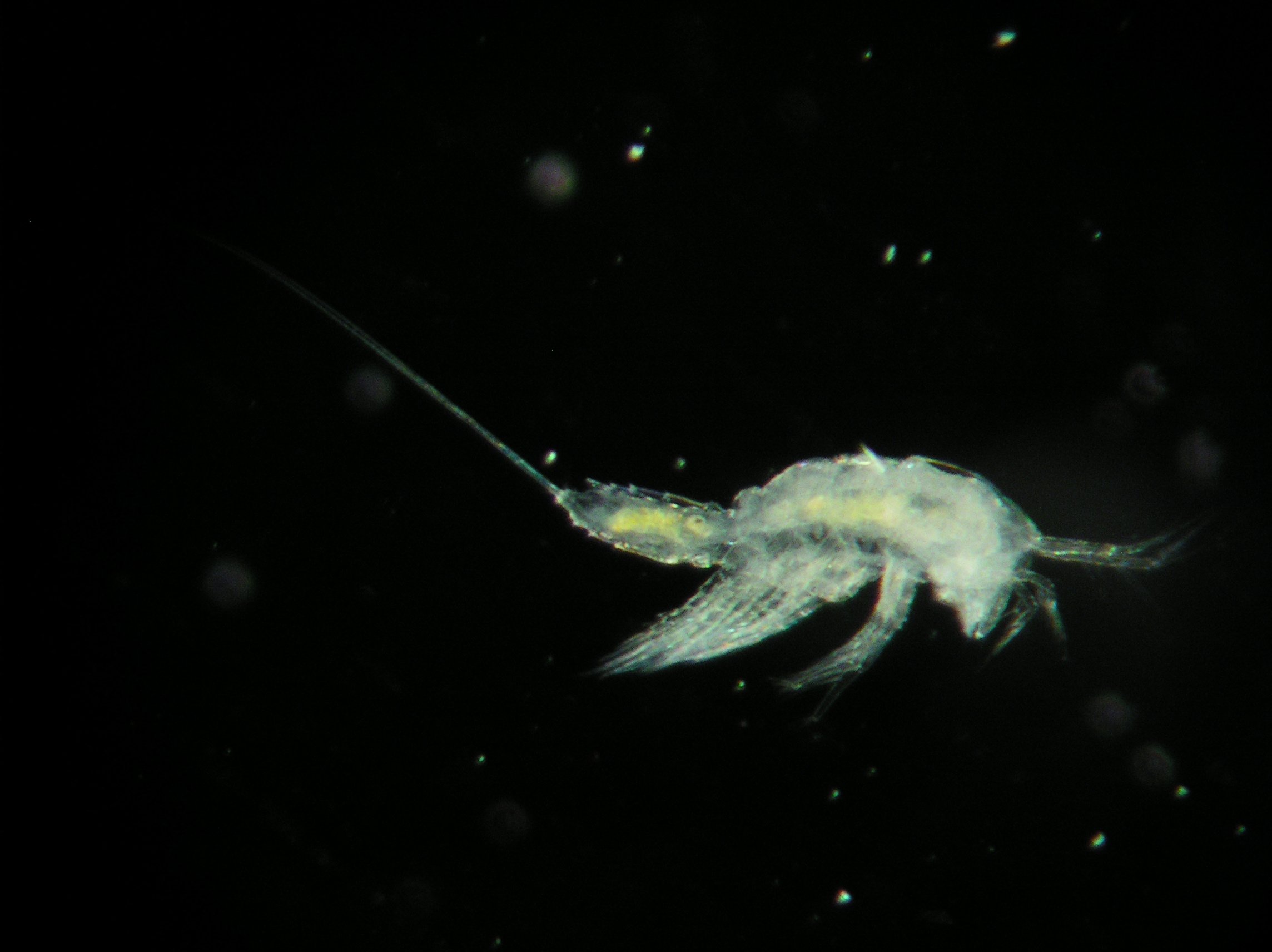
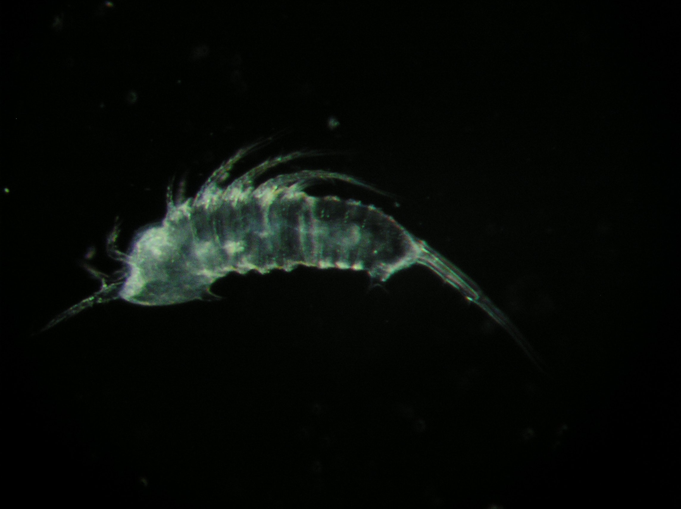 Surprisingly, copepods turned up in the traps—very well preserved ones that had apparently been alive until they met their demise in the formalin layer. Here are a few photos of the animals, as they look through the microscope. Laurence, the only member of the LOV staff who regularly works on animals that live on the sea floor (the place is otherwise a hotbed of plankton specialists), could tell that these copepods were not typical of the planktonic ones known from the area and suspected that she had found the first evidence of deep-sea emergence. David studies emergence—the strange tendency of some copepods that normally live in the sediment to emerge periodically into the water column—in shallow water; he's working out which species do it and which don't, and why they should do something so apparently maladaptive.
So they got to talking. The copepods would have to be identified before anyone could work out where they came from and what they were doing in the traps—were they mud dwellers? bottom crawlers? open-water swimmers? plankton, after all, of some strange new type? Laurence usually treats the bottom-dwelling copepods as a group, being interested in their collective influence on food chains, energy flow, etc., so didn't have the expertise to sort out the various species. David did, but had never worked on Mediterranean material of copepods that spent any time that far off the bottom.
David brought along his drawing tube—a microscope attachment that superimposes the image seen through the microscope and an image of the observer's hand, pencil, and paper, so that the observer can draw the image more or less by "tracing" it. (Not as easy as it sounds!) He and Laurence started out by combing the lab for a microscope with sufficient resolution that would also accept the drawing tube. They had reached the point of discussing makeshifts, when they happened to come by my office to say hi and instead said, "What's this under the cover? Look, it's already got a drawing tube on it!" Next, they spent several afternoons very, very carefully extracting the minute copepods from the eensy little vials in which they had been stored and making sure they were securely ensconced on a series of color-coded microscope slides, in Petri dishes for easy transport. Then, each afternoon, David would show Laurence how to mount a whole specimen or dissect one (remember that the larger individuals are the size of cornmeal and that each one has many, many tiny legs, each of which has many, many even tinier hairs, all of which are important to the identification) and then draw it. Fortunately, she already draws really well, but as I said, this drawing business is harder than it looks. The basic drawing is just the body, in profile, without the legs, but you're working with a specimen that's covered with legs (not to mention bacteria, diatoms, and bits of junk), and you can't see all three dimensions at once but have to focus up and down to see the details, and it's all nearly transparent, so it's not easy to tell what's in front of what. Each drawing took most of a day, and once we went home to dinner, Laurence would stay on to do her own drawing for comparison with David's the next day, and many's the time she would wail, "How can you see those details?! There's too much stuff in the way!" and David would say that maybe 30 years of practice had something to do with it. Anyway, they would spend some time every afternoon separating all the specimens into species, and each time they came to a new species they hadn't seen before, they would have to do the drawings of it. While one drew, the other worked on keying out the new beast in the work known as "Lange," a huge 1949 set of keys to harpacticoid species, all written in German by a scientist of that name. Copies are rare and unwieldy, so Laurence spent hours at the library last summer making a photocopy, which she bound in three volumes, each a good 5 inches thick. I think that, as of now, perhaps two of the species have proved to key out to something known—the rest are probably new to science.
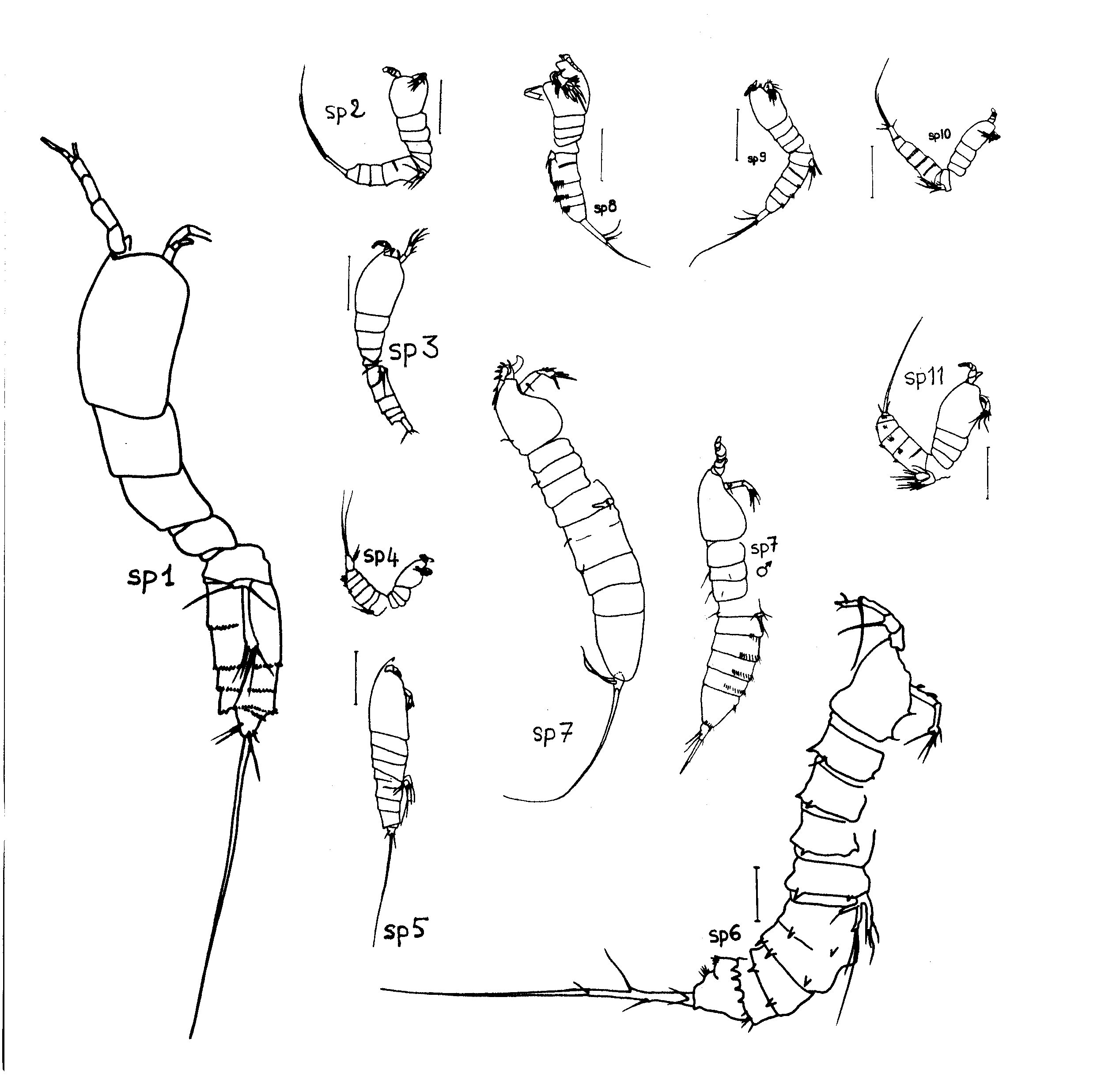 The group of drawings shown here was assembled when they had drawn 11 species. Each drawing was originally the size of the whole sheet of paper, i.e., as large as it could be and still fit within the field of view of the drawing tube. They were careful to include a scale bar in each drawing (representing 100 microns = 0.1 mm) so that the drawings could later be reduced on the photocopier until all were at the same scale. As you cn see, some species are much larger than others. All the individuals in this group of drawings are female, except that both sexes of species 7 are portrayed because they are so different.
David and Laurence just had time to get through all the specimens before we left Villefranche. Now, they're applying what they know about harpacticoid morphology and its correlations with different life styles (e.g., some animals have digging appendages, others appendages obviously adapted for swimming, others walking legs), what they know about where the probable known relatives of the unknown species live and how, and what is known about conditions at the site where the traps were deployed to try to formulate hypotheses about what the heck these animals were doing in the traps! Stay tuned; the work is still in progress.
previous entry List of Entries next entry