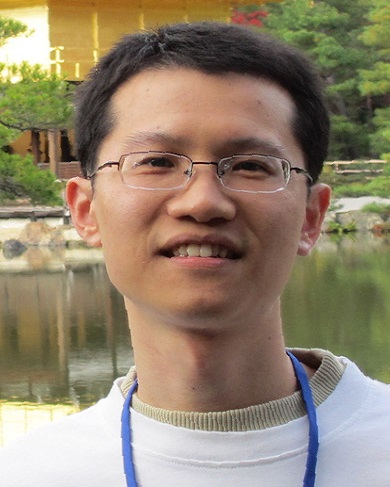
Biological Science Faculty Member
Dr. Guangxia Miao
- Office: 357 Biology Unit I
- Office: (850) 645-4065
- Area: Cell and Molecular Biology
- Lab: Biology Unit I
- Lab: 330
- Fax: (850) 645-8447
- Mail code: 4370
- E-mail: gmiao@bio.fsu.edu

Assistant Professor
PhD, Kobe University, 2015
Graduate Faculty Status
Research and Professional Interests:
In the intricate dance of life, cells are dynamic performers, constantly forming and breaking connections with one another. This dynamic interaction is pivotal during natural growth and in the occurrence of diseases. Imagine cells as dancers moving harmoniously in clusters, strands, sheets, and streams, navigating through a shifting environment and interacting with the surroundings and each other. Grasping and managing how cells behave under normal circumstances is fundamental for advancing fields like tissue engineering and regenerative medicine, which hold the promise of healing damaged tissues and organs. Moreover, this understanding is crucial as cells can, unfortunately, utilize their normal mechanisms for detrimental purposes, such as in cancer. Specifically, cancerous cells can exploit these mechanisms to spread – a process known as metastasis – posing significant challenges in cancer treatment. In contrast to stunning advances in uncovering the genetic control of primary tumor growth, the mechanisms underlying metastasis remain mostly elusive. Metastasis is a complex, multistep process that includes detachment of cells from the primary tumor, migration through neighboring tissues and the circulation, and attachment at new sites.
In the study of cell movement, much of attention has been focused on observing how cells traverse from one location to another. However, there remains a substantial gap in the understanding of how cell collectives break away from their initial neighbors in the process of delamination. Even less is known about how cells make new connections upon arrival at their ultimate destination. To be concise I name this process neolamination.
My lab established an in vivo model, the border cells in the Drosophila ovary, to study delamination and neolamination in collective cell movements. We deploy the powerful Drosophila genetics toolkit and organ culture methods to carry out high-resolution live imaging and optogenetics.
To date, the spread of tumor cells through the body, known as metastasis, continues to pose significant challenges in cancer treatment. This process begins with the detachment of tumor cells from the original site and concludes with their reattachment at a new location, mirroring similar phenomena observed during normal tissue development. My lab is focused on utilizing a robust experimental model I have developed to explore the cellular and molecular mechanisms driving these essential cellular behaviors.
Our long-term goal is to construct a conceptual model delineating how border cells interpret and respond to a myriad of external signals to facilitate collective delamination and neolamination. While our objective is not to replicate the entire metastatic process, we are keen on isolating and modeling individual steps to acquire profound mechanistic insights. We foresee that a more in-depth understanding of these processes will illuminate not only other normal morphogenetic events but also the behaviors of tumor cells. Ultimately, we aim to uncover fundamental principles that could significantly influence tissue engineering and identify novel therapeutic targets to address metastasis, the most lethal facet of cancer.
Lab website: https://gmiaoslab.wixsite.com/drmiaoslab
Selected Publications:
Miao, G., * Guo, L., and Montell, D.J.* (2022). Border cell polarity and collective migration require the spliceosome component Cactin. Journal of Cell Biology 211(7): e202202146.
Doi: https://doi.org/10.1083/jcb.202202146
Miao, G.*and Montell, D.J.* (2021). A thermo-genetics protocol for detecting gap junction channels in Drosophila egg chambers. Star Protocols 2, e100269 https://www.sciencedirect.com/science/article/pii/S2666166720302562?via%3Dihub
Miao, G., Godt, D., and Montell, D.J.* (2020). Integration of migratory cells into a new site in vivo requires channel-independent functions of innexins on microtubules. Dev. Cell 54, 501-515.e9. https://www.sciencedirect.com/science/article/pii/S1534580720305037
Highlighted in Dev. Cell Preview by Prof. Erez Raz https://www.sciencedirect.com/science/article/pii/S1534580720305529
Liufu, Z., Zhao, Y., Guo, L., Miao, G., Xiao, J., Lyu, Y., Wu, C.-I.* (2017). Redundant and incoherent regulations of multiple phenotypes suggest microRNAs’ role in stability control. Genome Res. 27, 1665-1673. https://genome.cshlp.org/content/27/10/1665.full
Miao, G. and Hayashi, S.*(2016). Escargot controls the sequential specification of two tracheal tip cell types by suppressing FGF signaling in Drosophila. Development 143, 4261–4271. https://journals.biologists.com/dev/article/143/22/4261/47483/Escargot-controls-the-sequential-specification-of
Miao, G. and Hayashi, S.* (2015). Manipulation of gene expression by infrared laser heat shock and its application to the study of tracheal development in Drosophila. Dev. Dyn. 244, 479–487. https://anatomypubs.onlinelibrary.wiley.com/doi/10.1002/dvdy.24192
Dong, B., Miao, G., and Hayashi, S.* (2014). A fat body-derived apical extracellular matrix enzyme is transported to the tracheal lumen and is required for tube morphogenesis in Drosophila. Development 141, 4104–4109. https://journals.biologists.com/dev/article/141/21/4104/46365/A-fat-body-derived-apical-extracellular-matrix
