 |
| BOT 3015 Final Exam Review | ||
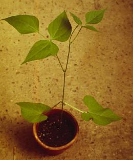 |
This bean seedling shows a pair of simple leaves as the lowest node (i.e., leaves are opposite). At the next node, only one leaf is present (i.e., alternate position); this leaf is compound--that is it has three leaflets. | |
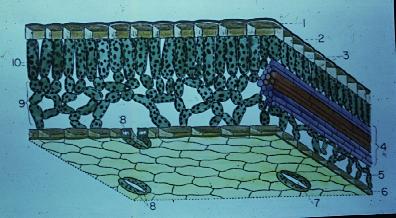 This leaf section shows epidermis (1) with stomates (7, 8) on lower epidermis. The mesophyll includes palisade and spongy parenchyma. The vascular bundle (vein, 4) includes xylem and phloem. |
||
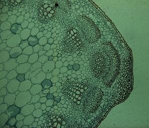 |
This stem cross section shows epidermis on the right side, followed by several cell layers of cortex (some collenchyma and some parenchyma), next is a series of vascular bundles (about five shown here) that form a ring, and finally the large parenchyma cells to the left of the picture constitute the pith. This is a dicot stem with no (or very little) secondary growth. | |
 |
This stem cross section shows several vascular bundles (of varying sizes); note that they are positioned out in the cortex and also deep into the pith region. This is a monocot stem (greenbrier). | |
 This stem has secondary growth (13 is secondary phloem; 14, secondary xylem). The "orange" row of cells (8) is a wood ray. 12 is the cortex, and 15 is the pith. This, of course, is a dicot stem. |
||
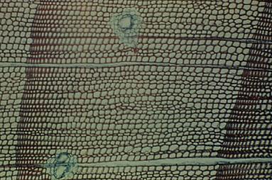 The transection (cross section) is of pine wood, showing parts of three annual rings. The one complete growth ring in center of view has early wood to the right and late wood to the left. Pine, a gymnosperm, has no vessels or fibers in its wood. The wood rays are one cell wide and run horizontally across the image. |
||
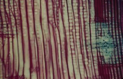 The radial section of pine wood shows the elongate tracheids vertically; the circular areas on the tracheids are pits. The darkly stained tracheids are part of the late wood, with tracheids of the early wood to the left. The "greenish-blue" cells to the right represent a wood ray as it typically appears in radial view. |
||
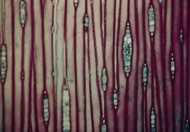 This tangential section of pine wood shows the same vertical orientation for the tracheids, but now the wood rays are seen as one cell wide and from three to ten cells tall. The one wood ray appears wider because it has a resin canal in it. |
||
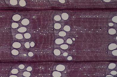 This transection of oak wood (a dicot) has parts of three growth rings. Large vessels are in the early wood, with much smaller ones in the late wood, thus the wood is called ring porous. Several uniseriate (one- cell wide) wood rays and two large, multiseriate wood rays are seen running horizontally. |
||
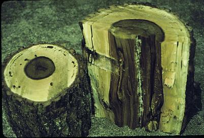 These cut logs of a dicot tree show the centrally located heartwood (darkly colored) and the lighter colored, outer sapwood near the cambium and the bark. |
||
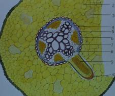 |
This chart shows a dicot root without secondary growth. The extensive yellow area is the cortex. Area 4 is the endodermis, and 5 is the pericycle. The dark "X" composed of larger cells is the xylem; phloem is orange (6). A branch root is coming from the pericycle (9). | |
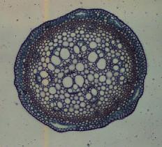
.jpg) This dicot root is said to be tetrach (four xylem poles oriented radially). The air passages in the cortex region indicate the species is adapted to a wet habitat. The absence of pith tells us this is a root rather than stem tissue. |
||
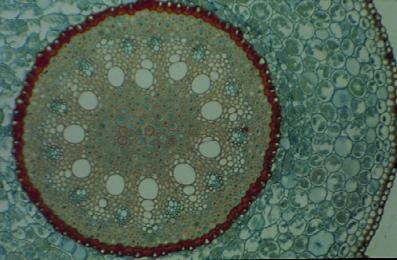 This monocot root shows numerous radially aligned rows of xylem and phloem (polyarch) which distinguishes is from a dicot root. Note the thick-walled cells (dark red) of the endodermis region. Dicot roots often have secondary growth; monocot root rarely have secondary growth. |
||
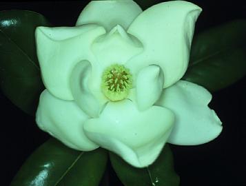 |
This Magnolia flower has three whorls of three petals each, many stamens, and many separate pistils. These features suggest to us that the species is a "primitive" angiosperm. They are pollinated by beetles which are relatively primitive insects, i.e., co-evolution. | |
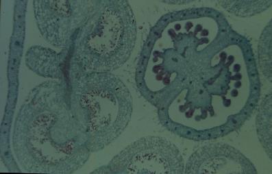 This cross section shows an anther on the left with four microsporangia containing pollen grains; on the right is an ovary with two internal chambers (said to be "axile" placentation). The several stubby, darkly-stained, finger-like projections are the ovules (young seeds). This flower came from a foxglove, the plant that gives us the heart medicine digoxin. |
||
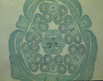 |
This view is a lily flower showing sepals, petals, six pollen-bearing anthers, and a centrally positioned ovary that has three chambers wherein the stubby, darkly-stained projections are the developing ovules. Flower parts in 3's suggests this is a monocot. | |
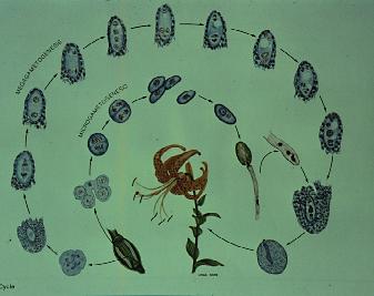 |
This lily represents the life cycle of an angiosperm. The inner circle shows an anther, pollen grains, one with a pollen tube delivering the sperm. The outer circle shows an ovary, then a megaspore mother cell undergoing meiosis to produce 4 megaspores, which in turn produce the 8-nucleate embryo sac (the mature megagametophyte), surrounded by integuments, ready for fertilization--then ultimately a seed with embryo. | |