bsc3402l\spec.html
Spectrophotometry
Spectrophotometry is one of the most useful tools available to the biochemist. It offers a high degree of precision, sensitivity, and accuracy. In addition, it is inexpensive and applicable to the measurement of a variety of substances. In this brief chapter, we will learn the principles of spectrophometry and how to use this technique with our laboratory instruments.
Principles
Organic compounds that absorb light are pigments. A ready example is chlorophyll itself, which absorbs light--as mentioned earlier--in the blue and red region of the visible light spectrum. For this reason, leaves are typically green (because they reflect green). If 5 g of leaf is extracted in an organic solvent, the extract (containing the solute chlorophyll) will be green. A leaf with a high chlorophyll content will yield a dark green extract, and a leaf with a low chlorophyll content will yield a pale green extract. Spectrophotometry is simply a means of measuring how densely green the solution is.
The absorption properties of an organic substance are constant, wherever.1 A spectrophotometer is an instrument that contains (a) a light source(s), (b) a means of isolating a particular wavelength band of the light source, (c) a sample holder, and (d) a device to measure light intensity. To measure the chlorophyll content of the extracts above, we would select a proper wavelength (say, 649 nm for chl b in 80% acetone2) on the spectrophotometer. Selection of wavelength is important because an extract or any solution may contain many compounds that absorb light. For the sake of the argument, say that your extract contains also carotenoids (as it would); carotenoids absorb in the roughly 450-540-nm region of the spectrum. Carotenoids are transparent in the > 600 nm region of the spectrum. So selecting 649 nm prevents carotenoids from interfering with your measurement of light absorption. Of course, this same argument would apply to other potentially interfering substances in the extract, but you must be alert to the possibility that some nonchlorophyll substances in the extract would absorb light at 649 nm, so a
_________________________
1 This statement, of course, is based on the presumption of constancy of the system. E.g., in the extraction of chlorophyll, one must take reasonable precautions to avoid oxidation. The solvents must be similar in compared solutions--the absorption maximum will be somewhat different in different solvents such as ether or pyridine.
2 Although you select "649 nm," the light that passes though the sample will not be exactly and absolutely 649 nm. On a clinical or student instrument such as the spec 20, "649 nm" means that about 75% of the light will be between 629 and 669 nm. On routine research instruments, the wavelength selection is much better, say ± 1-2 nm.
single wavelength measurement provides for only a somewhat equivocal conclusion. For each particular substance, one hopes to devise a means of reducing the ambiguity of the measurement. In the case of chlorophyll, the prudent investigator would typically take a measurement also at 700 nm, where chlorophyll does not absorb to an appreciable degree and which is fairly close to 649 nm. (Particulates in the extract would reflect light at 700 nm, so this is a good control to run with dilute solutions of chlorophyll for this additional reason also.) Next, we would ensure that the proper lamp is on and warm. Then we would blank the instrument. The electrical output of the light measuring device depends on several parameters, such as the "dark current" (electronic noise) of the instrument and the intensity of the illuminating beam. To blank the instrument, place some absolutely constant solution in the light path. In the visible region of the spectrum, water is an ideal choice. Water is readily available, cheap, and will not change. With no light striking the light-measuring device, set the instrument to 0% transmittance. With the cuvette containing the water in the light path between the light source and the measuring device (typically, a photomultiplier tube), set the instrument to 100% transmittance. This is a matter of changing the "gain" of the electronic circuit. Of course, all subsequent measurements must be made with the same type of cuvette, &c. With the instrument properly set up, place the sample in the cuvette and take a measurement. Proper controls must also be run; in this case, a sham extract--containing no leaf material--must be read because components of the extract cocktail itself may absorb at 649 nm. It would be even better if we could read a single tube and selectively convert chlorophyll to a colorless compound and take a second reading. The difference between the first and second reading would be a precise measurement of light absorption by chlorophyll in the extract.
The spectral properties of a substance may change, depending on the chemical environment and modifications to the molecule. In some cases, "simply" changing the pH will convert a colored substance to a colorless one. In other cases, the oxidation state of the molecule will cause dramatic changes in the spectral properties. The relevant example here is NADP. The oxidized form, NADP+, does not absorb light at 340 nm, and the reduced form, NADPH, does. Your assay,
1,3-diPGA + NADPH ® PGal + NADP+
will therefore result in a loss of light absorption at 340 nm. The task, then, is to relate the rate of loss of light absorption to the rate of disappearance of NADPH. Enzyme activity, by definition, is the rate of substrate consumption per unit time. (As you will learn in another section, enzyme specific activity--i.e., activity put on some unit tissue basis--is more often the desired quantity.)
Given constant conditions, as alluded to (and explained again in the section on calculation of enzyme activity), the absorption by a particular species at a particular wavelength is constant. Although the instrument itself is essentially measuring transmittance (the percentage of incident radiation that passes through the sample), it is not convenient to use transmittance because the concentration of the absorbing species is not linear with transmittance. However, a simple mathematical conversion, to Optical Density (O.D.) or Absorption, creates a linear relationship:
O.D. = - log (T)
An infinitely concentrated solution will have a T = 0% and O.D.= infinity. A "solution" with no solute will have a T = 100% and O.D. = 0. Although research grade instruments can read very dilute and very concentrated solutions, we will be using as a starting point the center of the scale. You will read O.D. directly off the instrument; express your O.D. as A340.
The so-called x mM (say "millimolar extinction coefficient") at 340 nm with a 1-cm light path for NADPH is 6.27. In other words, a 1 mM solution of NADPH will have an O.D. of 6.27 at 340 nm and with a 1-cm light path. Since the O.D. and concentration are linear, a 0.5 mM solution will have an O.D. of 6.27 x 0.5 &c. You may wish to remember that and O.D. of 1.00 corresponds to a 160-m M solution. In your assay system, you will start with a solution containing NADPH, and PGal DH will catalyze NADPH oxidation with time. By using the extinction coefficient, you will calculate the rate of loss of NADPH.
Practical Aspects
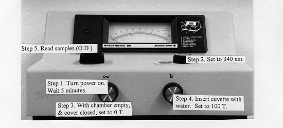
This section provices simple straight-forward directions for use of the spec20. Please refer to the photograph and follow the label directions.