The Nerve Impulse Seen from Outside
Dexter M. Easton July 2000 ©
Next topic Previous topic Table of Contents
Topic 17: Longitudinal currents at the recorded end, cut and healed
During dissection, the nerve must be cut across, and the membrane barrier between inside and outside of each nerve fiber is breached. There exists, in effect, a "short circuit" between the positive outside and the negative inside of the nerve fibers. Within half an hour, the cut ends "heal over," and the high-resistance membrane is reestablished. In that circumstance, the resting voltage between the recording electrodes is zero. There is no longitudinal current, because there is no longitudinal potential gradient. A gradient is detected when an impulse invades the nerve end within the recording tube. The record of the impulse is diphasic: an initial upward deflection due to the leading currents, followed by the downward deflection due to the trailing currents.
The diphasic (positive, then negative) form of the AP is obtained only after the nerve has been "resting" in a compatible solution for half an hour or more after the ends have been cut. During this time, the integrity of the cut membrane is reestablished.
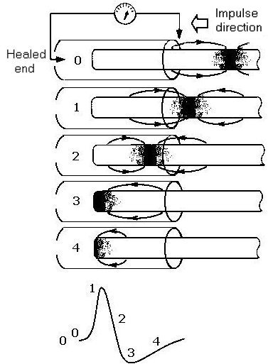
In Fig. 17A, as the impulse moves along, the direction and density of longitudinal currents determines the amplitude of the action-potential signal. The figure shows "snapshots" relating the signal and the position of the currents in the recording tube. At (1) the detected currents are toward the right, yielding the upward (+) signal reflecting the leading currents. At (2) oppositely directed trailing currents cut the signal down, and at (3) only the trailing currents are present, and these die out at 4.
Figure 17A. The diphasic action potential and the healed nerve end.
If the end of the nerve has been freshly severed, a voltage difference, the injury potential, can be measured along the injured stump. A simple equivalent circuit shows the relation of this potential to the resting membrane potential. Application of Ohm’s law shows why the injury potential is only a small fraction of the total available resting membrane potential.
At the cut end the + charges on the outside of the membrane have easy access to the negative countercharges inside. Through this region of low resistance, current (injury current) will flow from the outside toward the inside via the cut end. The depolarizing gradient, greatest at the end, declines approximately exponentially with distance away from the level of the cut.
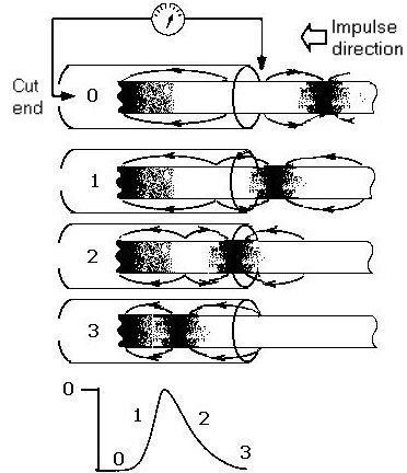
Figure 17B. The injury (cut end) potential and the monophasic action potential.
Fig. 17B (like Fig. 17A) shows "snapshots" relating currents and the action potential. Initially, at (0), there is a steady negative deflection due to injury currents directed toward the left within the recording tube. As the impulse moves toward the left, the leading currents, directed toward the right, drive the signal upward (1). This effect is counteracted by the oncoming trailing currents (2), and as those die out, the injury currents again dominate (3).
Next topic Previous topic Table of Contents